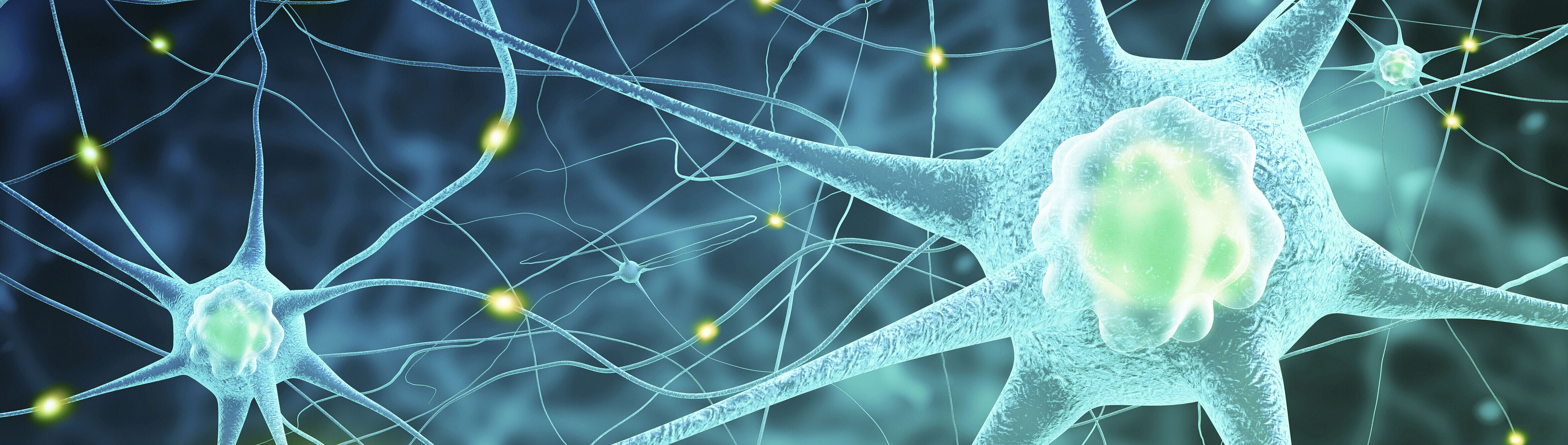
From Macroscopy to Histology: Organization of the Central Nervous System
Medical Winter School - MedWin 2026
From macroscopy to histology: organization of the central nervous system
The University of Rostock is excited to welcome all participants and speakers to our Medical Winter School!
MedWin 2026 offers an opportunity to experience a fascinating academic and cultural program with the University of Rostock from March 21st to March 29th 2026. Feel free to reach out for any questions regarding MedWin 2026: medwin.io@uni-rostock.de
Program Overview
Duration: 6 days
Location: Institute of Anatomy and selected research laboratories, University Medical Center Rostock and University of Rostock
Core Components
• Human brain dissections supervised by anatomical experts
• Histology sessions using high-resolution microscopy and digital imaging systems
• Lectures and seminars on major CNS diseases (e.g., multiple sclerosis, Parkinson’s disease, stroke)
• Panel Discussion with international experts (topic tbd)
• guest lectures by international experts in the field of neurosciences
Additional Program Highlights
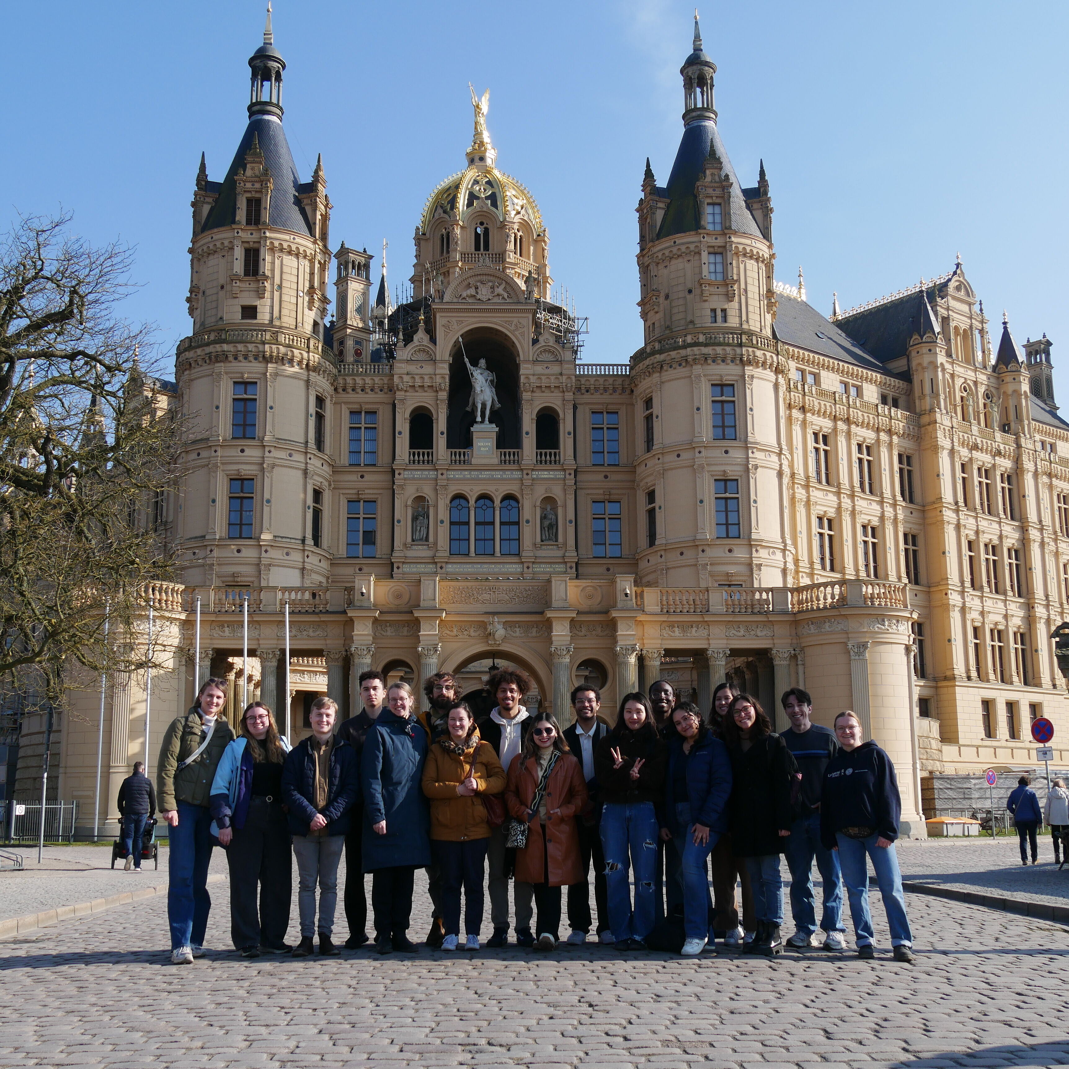
• A full-day excursion to Schwerin, the capital city of Mecklenburg-Vorpommern
• Presentation “Report of a Ship’s Doctor” by Dr. Gerd Schroeter at the officer's mess on former GDR freighter MS “Dresden”
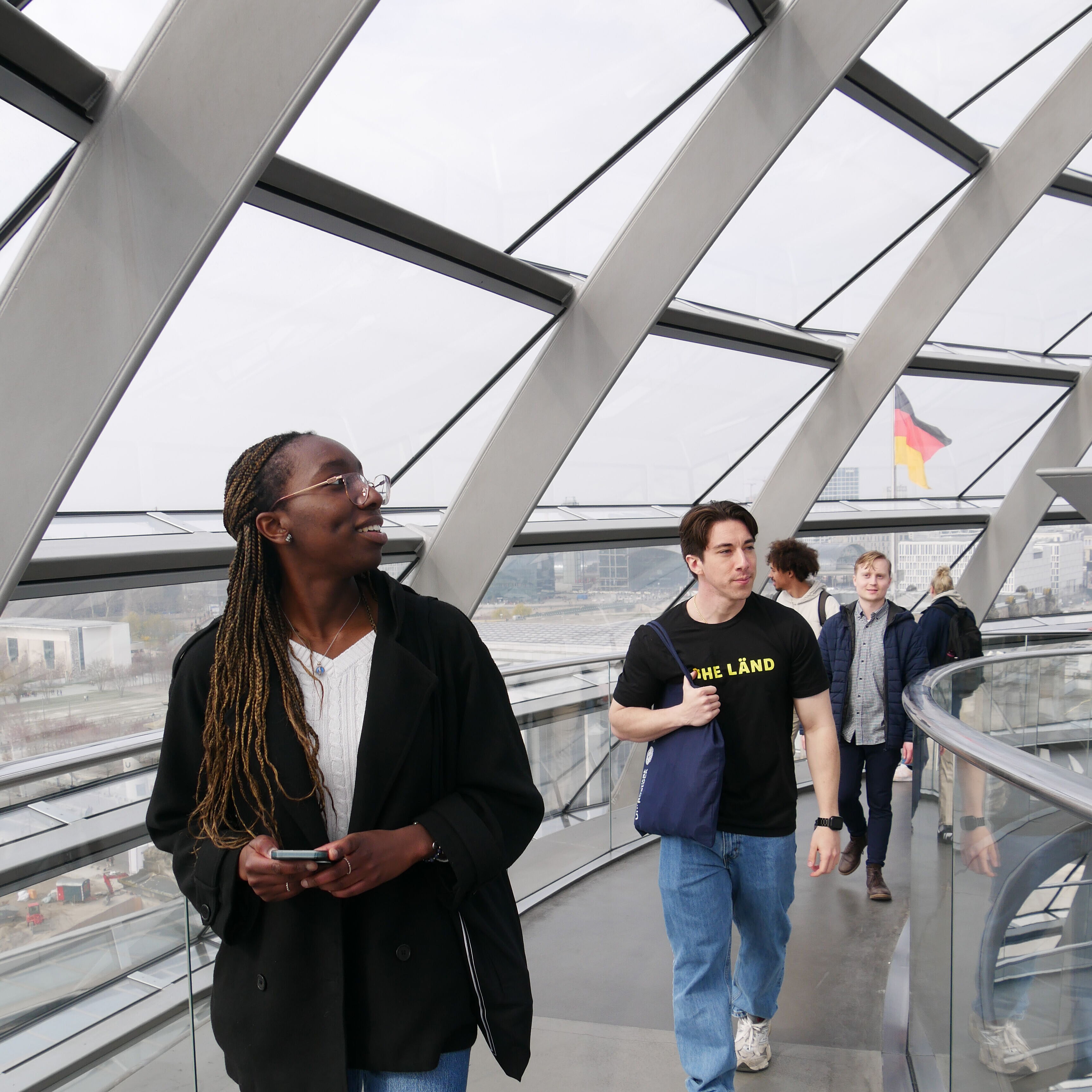
• Trip to Berlin including visits to the Charité Museum and the German Parliament, plus free time to explore the city independently
Specific Aims
• provide deep insight into the structural and functional organization of the CNS
• foster neuroanatomical knowledge through human brain dissection
• introduce students to neuropathological concepts and neurological disease patterns
• promote international exchange among students in medicine and related disciplines
Quick Facts
| Date | March 21 to 29, 2026 |
|---|---|
| Venue | University of Rostock, Medical School |
| Target group | Students with medical backround |
| Language | English |
| Travel Grant | 300€ per participant |
| Credits | Certificate |
Contact:
Dr. Christoph Rothenbuecher (he/him)
Short-Term Programs Coordination
medwin.io@uni-rostock.de
+49 381 498 9806
Fone Hours: Monday – Friday 9 – 12
Room 116 – Friedrich-Engels-Platz 6
18055 Rostock – Germany

Please note that this programm will be subject to changes!
We are honored to welcome these speakers to MedWin 2026
Karen Tashima
Professor of Medicine at Brown University
Karen Tashima is a professor of medicine in the Division of Infectious Diseases at the Brown University in the United States. She is the Director of the HIV Clinical Studies and Clinical Research Site Leader of the AIDS Clinical Trials Unit at The Miriam Hospital. Dr. Tashima is the study chair of an ACTG study evaluating the effectiveness of a new strategy to treat HIV-infected persons with drug resistant virus. In 2005, she received the HIV Leadership Award as Outstanding HIV/AIDS Clinical.
Seema K. Tiwari-Woodruff
Professor for Biomedical Sciences and Director of Biomed Graduate Program at UC Riverside
Seema K. Tiwari-Woodruff is a Professor of Biomedical Sciences in the School of Medicine at the University of California, Riverside. She is a recognized leader in multiple sclerosis research, where her work focuses on understanding mechanisms of neurodegeneration and inflammation and developing strategies to promote neural repair and remyelination. In addition to leading an active research program, Dr. Tiwari-Woodruff serves as Director of the Biomedical Sciences Graduate Program at UCR, guiding graduate education and mentorship across the biomedical sciences. She is also deeply committed to education, teaching both first-year medical students and graduate students.
Alina Blusch
Associate researcher at the Department of Experimental Medical Science, Lund University
Alina Blusch is a researcher at Lund University in Sweden. Dr. Blusch completed her PhD in neuroscience at the International Graduate School of Neuroscience, Ruhr University Bochum. Her research focuses on inflammation in Huntington’s disease, with the aim of elucidating immune-related mechanisms contributing to disease progression and neurodegeneration.
Michael Punsoni
Director of Autopsy Services, Director of Ophthalmic Pathology and Associate Professor at Brown University
Michael Punsoni is board certified in Anatomic and Clinical Pathology, and Neuropathology by the American Board of Pathology. He has received several teaching awards for his commitment to education, including the Ronald A. DeLellis Mentoring Award from Brown University. Dr. Punsoni has lectured nationally and internationally on a variety of topics related to brain tumors, dementia, ophthalmic pathology and autopsy-related cases. He has been involved in multiple grant-supported research projects, and is dedicated to investigating the pathophysiology of brain tumors and neurodegenerative disease-related biomarkers.
Ursula van Rienen
Professor for Electromagnetic Field Theory / former Spokesperson of the Research Center ELAINE
Prof. Dr. Ursula van Rienen works at the Institute for general electrical engineering. Her main research topics are the modelling and simulation of questions from accelerator physics and accelerator technology, especially beam dynamics and the fields in high-frequency structures. She is former head of the DFG Collaborative Research Center for Electrically Active Implants (ELAINE) focusing on specifically implants employed for the regeneration of bone and cartilage, and implants for deep stimulation to treat movement disorders.
Prof. Dr. Markus Kipp
Director of the Institute of Anatomy at the University medical center Rostock
Since 2018 Prof. Dr. Kipp is Director of the Institute of Anatomy. He and his research group are interested to understand the physiology and pathology of the axon-oligodendrocyte-myelin unit. Particularly, they are interested to understand to what extend stressed oligodendrocytes regulate peripheral immune cell recruitment, and by which neuroanatomical pathways peripheral immune cells gain access into the CNS parenchyma. To address their scientific challenges they use state of the art techniques such as design-based stereology, a set of behavioral analyses such as high speed ventral plane videography, or positron emission tomography/computed tomography. Whenever possible, they compare their pre-clinical results with post-mortem samples, obtained from MS patient donors.
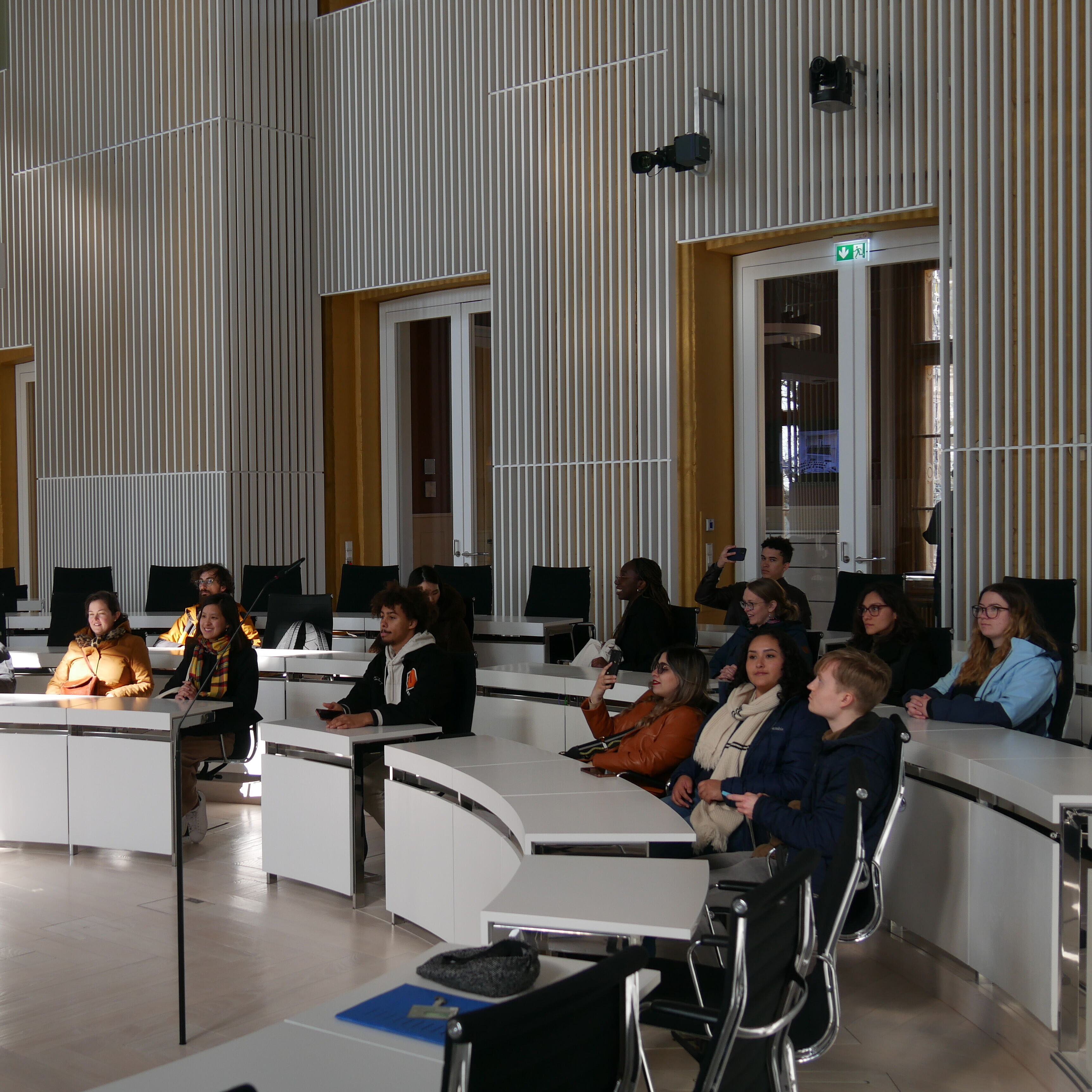
Impressions of last year's MedWin
Copyright by Lena Breuer